Dermatology Terminology
Ontario Association of Veterinary Technicians. (2022). [Dermatology]. Brought to you by OAVT Seminar.
MUCINOSIS
- Folds in skin, normal folds due to breed
EDEMA

Facial edema → “Tragic expression”
Mixed edema → may include tail base edema (1)
First action: Check thyroid function
Tail base edema (“jelly butt”) → indicative of fluid accumulation (1)
Erythema (skin redness) → chronic inflammation → post-inflammatory hyperpigmentation (2)

Persistent inflammation → Lichenification (thickened, leathery skin) (3)
Lichenified skin → ideal surface for secondary infections
HYPERKERATOSIS

Thickened Skin & Nasal/Foot Pad
Thickened areas are often the first visible sign of systemic or internal disease, especially on nasal and foot pads.
Indicator of other diseases:
- Canine distemper (foot pads)
- Leishmaniasis
- Pemphigus (most common autoimmune skin disease)
- Zinc-responsive dermatosis (nutritional absorption issue)
- Hepatocutaneous syndrome / Superficial necrolytic dermatitis (liver-related)
If all other disease have been ruled out along with other diagnosis by process of elimination the case can be an Idiopathic conditions:
-
Nasodigital hyperkeratosis- common with aging
-
Hereditary nasal parakeratosis (common in Labrador Retrievers)
CUTANEOUS ATROPHY

Thinning Skin & “Old Lady” Skin in Dogs
-
Skin changes: Thinning, folding, or “chicken skin” appearance.
-
Causes:
-
Long-term steroid use
-
Cushing’s disease (excess corticosteroid production)
-
-
Associated lesions: Papules and redness may develop due to fragile skin and inflammation.
Comadomes(blackheads/blocked hair follicles)
Abnormal thickening → disrupts normal skin function and barrier.
Lifted skin → often a collarette, marking the footprint of a previous pustule
Pustules

Scaleing

- Increased scaling very fine and varied location in Hypothyroid
- Large flakes Ichthyosis (dry skin) Golden retrievers overrepresented
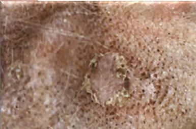
Crusts

- Some juicing
Gold crust commonly seen with :
( 1)Immune mediated( Pemphigus)
or
Sarcopties (2)

Silver crust --> Do Skin Biopsy
- Epithelial Tropic Lymphoma (Cancer/Cutaneous T cell lymphoma)
- Most are Euthanized as symptoms (Severe itchiness pruritis) are rarely put under control
Vesicles

Blisters
Check in the mouth oral lesions, prepuce, vulva =>Usually seen in immune mediated or autoimmune diseases
WHEELS/HIVES/Urticaria

Draining tracts

- Interdigital cyst
- Foreign body reaction
- Sterile abscess


Comments
Post a Comment